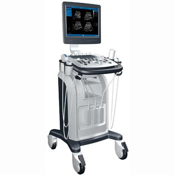
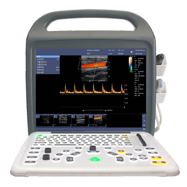
Color Doppler ultrasound diagnostic instrument
MIANYANGXIANFENGMEDICALINSTRUMENTCO.,LTD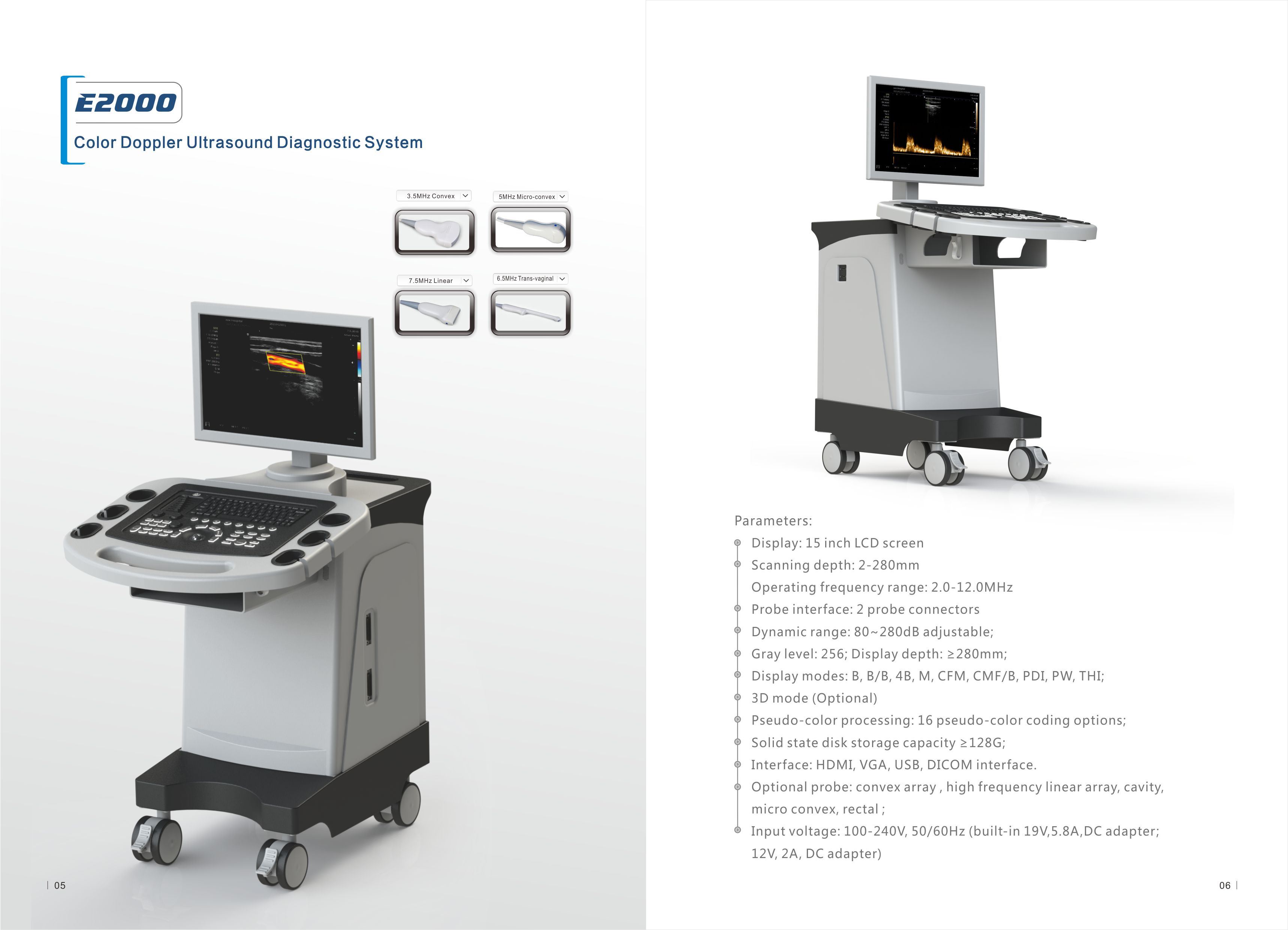
The supplier hasn’t listed a price. We can get their current prices and MOQ.
Verify from $29 · written report in 3 business days · 100% refund if not satisfied
Color Doppler ultrasound diagnostic instrument
MIANYANGXIANFENGMEDICALINSTRUMENTCO.,LTDThe supplier hasn’t listed a price. We can get their current prices and MOQ.
Verify from $29 · written report in 3 business days · 100% refund if not satisfied
About this product
- Noise level: Other
- Parts: Other
- Exports to: Africa, Middle East, Southeast Asia, South Asia, Central Asia, USA and Canada, Latin America, Russia
About this product
- Noise level: Other
- Parts: Other
- Exports to: Africa, Middle East, Southeast Asia, South Asia, Central Asia, USA and Canada, Latin America, Russia
Product details
| Noise level | Other |
|---|---|
| Parts | Other |
Product details
| Noise level | Other |
|---|---|
| Parts | Other |
From the supplier
E2000 Brief introduction
Display: 15 inch LCD screen
Scanning mode: electron convex array, high frequency linear array, cavity, micro convex, rectum;
Image mode:
R B, B+B, 4B mode
R B+M mode
R CFM color blood flow pattern
R B+CFM mode
R PDI energy mode
R PDI+B mode
R PW mode
R B tilt mode (Note: B- tilt only applies to high-frequency linear probes less than or equal to 40mm.)
R 3D mode (Optional)
Scanning depth: 2-280mm
Operating frequency range: 2.0-12.0MHz
Probe interface: 2 automatic probe identification ports
Dynamic range: 80~280dB adjustable;
Display modes: B, B/B, 4B, M, CFM, CMF/B, PDI, PW, THI;
Application mode: abdomen, gynecology, obstetrics, superficial organs, pediatrics, urology, heart, blood vessels and other modes;
Imaging modes: full digital multi-beamforming, speckle noise suppression, tissue harmonic imaging, etc
Sound output: real-time display of mechanical index and thermal index, adjustable sound power and real-time display;
Gray level: 256;
Display depth: ≥280mm;
Pseudo-color processing: 16 pseudo-color coding options;
Gain adjustment: 8 segments TGC, B/M/D/C gain can be adjusted respectively, TGC curve can be displayed and automatically hidden;
Image processing: Level 5 image optimization, edge enhancement, frame average, Line average, focus optimization, noise suppression, Gamma correction, closing curve, contrast
Degree, brightness adjustable, up and down, left and right flip;
Automatic optimization function: built-in multiple check types, according to different check types, preset the best image check conditions, reduce the adjustment operation keys;
Measurement and calculation: B mode conventional measurement, distance, circumference, area, volume, Angle, ratio, and shorthand rate, M mode conventional measurement, woman
Department measurement, obstetrics measurement, cardiology measurement, urology measurement, PW measurement and other measurements.
Image annotation: alphanumeric input, adjustable annotation arrows, labels and comments, body markers, patient and hospital ids, etc.
Image storage: image storage, video storage. Movie playback, solid state disk storage capacity ≥128G;
Patient data: medical record management, report query and printing, image and video output (hard disk, USB, optional DVD-RW), built-in ultrasound workstation;
Report page system: automatic report generation system, and full-screen characters in Chinese and English editing;
Interface: HDMI, VGA, USB, DICOM interface.
Support probe: electronic linear array CT7.5L40GN, electronic convex array CT3.5C60GN, electronic cavity CT6.5C10GN, electronic slightly convex CT3.5C20GN
Electronic convex CT6.5C8015, electronic convex CT7.5C8020, electronic rectal CT6.0L8064
Input voltage: 100-240V, 50/60Hz (built-in 19V,5.8A,DC adapter; 12V, 2A, DC adapter)
Appearance size: 590mm×885mm×1205mmm (length × width × height)
Weight ≤ 18Kg
From the supplier
E2000 Brief introduction
Display: 15 inch LCD screen
Scanning mode: electron convex array, high frequency linear array, cavity, micro convex, rectum;
Image mode:
R B, B+B, 4B mode
R B+M mode
R CFM color blood flow pattern
R B+CFM mode
R PDI energy mode
R PDI+B mode
R PW mode
R B tilt mode (Note: B- tilt only applies to high-frequency linear probes less than or equal to 40mm.)
R 3D mode (Optional)
Scanning depth: 2-280mm
Operating frequency range: 2.0-12.0MHz
Probe interface: 2 automatic probe identification ports
Dynamic range: 80~280dB adjustable;
Display modes: B, B/B, 4B, M, CFM, CMF/B, PDI, PW, THI;
Application mode: abdomen, gynecology, obstetrics, superficial organs, pediatrics, urology, heart, blood vessels and other modes;
Imaging modes: full digital multi-beamforming, speckle noise suppression, tissue harmonic imaging, etc
Sound output: real-time display of mechanical index and thermal index, adjustable sound power and real-time display;
Gray level: 256;
Display depth: ≥280mm;
Pseudo-color processing: 16 pseudo-color coding options;
Gain adjustment: 8 segments TGC, B/M/D/C gain can be adjusted respectively, TGC curve can be displayed and automatically hidden;
Image processing: Level 5 image optimization, edge enhancement, frame average, Line average, focus optimization, noise suppression, Gamma correction, closing curve, contrast
Degree, brightness adjustable, up and down, left and right flip;
Automatic optimization function: built-in multiple check types, according to different check types, preset the best image check conditions, reduce the adjustment operation keys;
Measurement and calculation: B mode conventional measurement, distance, circumference, area, volume, Angle, ratio, and shorthand rate, M mode conventional measurement, woman
Department measurement, obstetrics measurement, cardiology measurement, urology measurement, PW measurement and other measurements.
Image annotation: alphanumeric input, adjustable annotation arrows, labels and comments, body markers, patient and hospital ids, etc.
Image storage: image storage, video storage. Movie playback, solid state disk storage capacity ≥128G;
Patient data: medical record management, report query and printing, image and video output (hard disk, USB, optional DVD-RW), built-in ultrasound workstation;
Report page system: automatic report generation system, and full-screen characters in Chinese and English editing;
Interface: HDMI, VGA, USB, DICOM interface.
Support probe: electronic linear array CT7.5L40GN, electronic convex array CT3.5C60GN, electronic cavity CT6.5C10GN, electronic slightly convex CT3.5C20GN
Electronic convex CT6.5C8015, electronic convex CT7.5C8020, electronic rectal CT6.0L8064
Input voltage: 100-240V, 50/60Hz (built-in 19V,5.8A,DC adapter; 12V, 2A, DC adapter)
Appearance size: 590mm×885mm×1205mmm (length × width × height)
Weight ≤ 18Kg
About the supplier
- ✓Exhibited at 5 recent Canton Fairs
- ✓Manufacturer
- ✓Founded 2005
- ✓Canton Fair Premium Supplier
- ✓National high-tech enterprise
Health & Recreation exhibitors show in Phase 3 of the Canton Fair, 31 October to 4 November 2026.
- Exhibited at 5 recent Canton Fairs
- Manufacturer
- Founded 2005
- Canton Fair Premium Supplier
More from MIANYANGXIANFENGMEDICALINSTRUMENTCO.,LTD


Similar products from other suppliers